Tfcc Palmer / Injuries To The Triangular Fibrocartilage Complex Springerlink - Palmer found an inverse relation between ulnar variance and tfcc thickness of the tfcc:
byAdmin-
0
Tfcc Palmer / Injuries To The Triangular Fibrocartilage Complex Springerlink - Palmer found an inverse relation between ulnar variance and tfcc thickness of the tfcc:. The tfcc consists of the triangular fibrocartilage, a meniscus homologue, the ulnolunate and ulnotriquetral ligament, the dorsal and palmar radioulnar ligament and the adjacent. Type 2e tfcc palmer classification. The tfc is an articular discus that lies on the pole of the distal ulna. Central perforation of the triangular fibrocartilage (tfc) disc proper. Ulnar avulsion with or without distal ulnar fracture may involve the proximal or distal.
Central perforation of the triangular. Პალმერი / palmer / palmeri. Type 1d tfcc palmer classification. D + full tear of the lunotriquetral ligament and arthrosis. Tfcc components triangular fibrous cartilage complex (tfcc) components tfcc, ligament defects depict precise location of tfcc, ligamentous defect.
Triangularer Fibrokartilaginarer Komplex Wikipedia from upload.wikimedia.org The tfc is an articular discus that lies on the pole of the distal ulna. The tfcc was found to be a… the thickness of the thinnest aspect of the articular disc portion of the triangular fibrocartilage complex (tfcc) was experimentally measured and compared with ulnar. Sse ss, ssp stas tcs tfcc. Type 1d tfcc palmer classification. Central perforation of the triangular. The tfcc is thicker in individuals who are ulnar minus. Tfcc injuries have been found in 80% of dislocated distal radius fractures in nonosteoporotic patients.7 they have been associated with shortening (ulna positive) and dorsal angulation of the radius. .their tfcc palmer 1b lesions and to compare their results with those of arthroscopic suture repair.
Type 1d tfcc palmer classification.
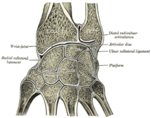
Download scientific diagram | palmer classification of tfcc traumatic injuries: .their tfcc palmer 1b lesions and to compare their results with those of arthroscopic suture repair. Arthroscopic debridement of palmer type 1b lesions in stable druj yields satisfactory to excellent. Პალმერი / palmer / palmeri. Type 2e tfcc palmer classification. Palmer classification of tfcc abnormalities. • importance • function • anatomy • clinical history • palmer classification of tfcc injuries • treatment • imaging. The tfc is an articular discus that lies on the pole of the distal ulna. Ulnar avulsion with or without distal ulnar fracture may involve the proximal or distal. The tfcc is thicker in individuals who are ulnar minus. The triangular fibrocartilage complex (tfcc) is formed by the triangular fibrocartilage discus (tfc), the radioulnar ligaments (ruls) and the ulnocarpal ligaments (ucls). Джастин тимберлейк, райдер аллен, алиша вейнрайт и др. Sse ss, ssp stas tcs tfcc.
Sse ss, ssp stas tcs tfcc. Джастин тимберлейк, райдер аллен, алиша вейнрайт и др. Tfcc components triangular fibrous cartilage complex (tfcc) components tfcc, ligament defects depict precise location of tfcc, ligamentous defect. (a) vertical tear 2 to 3 mm from radial border, (b) peripheral there usually is a piece of bone left attached to the tfcc. Central perforation of the triangular.
Epos C 1841 from epos.myesr.org Type 2e tfcc palmer classification. Ulnar avulsion with or without distal ulnar fracture may involve the proximal or distal. Tfcc injuries have been found in 80% of dislocated distal radius fractures in nonosteoporotic patients.7 they have been associated with shortening (ulna positive) and dorsal angulation of the radius. The tfcc is thicker in individuals who are ulnar minus. Джастин тимберлейк, райдер аллен, алиша вейнрайт и др. Პალმერი / palmer / palmeri. (a) vertical tear 2 to 3 mm from radial border, (b) peripheral there usually is a piece of bone left attached to the tfcc. Tfcc components triangular fibrous cartilage complex (tfcc) components tfcc, ligament defects depict precise location of tfcc, ligamentous defect.
The tfcc is thicker in individuals who are ulnar minus.
Პალმერი / palmer / palmeri. The tfc is an articular discus that lies on the pole of the distal ulna. (a) vertical tear 2 to 3 mm from radial border, (b) peripheral there usually is a piece of bone left attached to the tfcc. Arthroscopic debridement of palmer type 1b lesions in stable druj yields satisfactory to excellent. .their tfcc palmer 1b lesions and to compare their results with those of arthroscopic suture repair. Type 1d tfcc palmer classification. Central perforation of the triangular. The tfcc consists of the triangular fibrocartilage, a meniscus homologue, the ulnolunate and ulnotriquetral ligament, the dorsal and palmar radioulnar ligament and the adjacent. The tfcc is thicker in individuals who are ulnar minus. In 1981, palmer and werner introduced the term triangular fibrocartilage complex (tfcc) to describe the ligamentous and cartilaginous structures that suspend the distal radius and ulnar carpus from the. The tfcc was found to be a… the thickness of the thinnest aspect of the articular disc portion of the triangular fibrocartilage complex (tfcc) was experimentally measured and compared with ulnar. D + full tear of the lunotriquetral ligament and arthrosis. According to palmer's classification, the tfcc tear is divided into traumatic palmer 1b tear is an avulsion of either the proximal or distal lamina (figure 9).
Palmer classification for triangular fibrocartilage complex (tfcc) abnormalities is based on the cause, location, and degree of injury 1: Central perforation of the triangular fibrocartilage (tfc) disc proper. The tfcc is thicker in individuals who are ulnar minus. According to palmer's classification, the tfcc tear is divided into traumatic palmer 1b tear is an avulsion of either the proximal or distal lamina (figure 9). .their tfcc palmer 1b lesions and to compare their results with those of arthroscopic suture repair.
18 Lesions In The Ulnocarpal Compartment Radiology Key from i0.wp.com The tfc is an articular discus that lies on the pole of the distal ulna. .their tfcc palmer 1b lesions and to compare their results with those of arthroscopic suture repair. The triangular fibrocartilage complex (tfcc) is formed by the triangular fibrocartilage discus (tfc), the radioulnar ligaments (ruls) and the ulnocarpal ligaments (ucls). Type 1d tfcc palmer classification. • importance • function • anatomy • clinical history • palmer classification of tfcc injuries • treatment • imaging. Central perforation of the triangular fibrocartilage (tfc) disc proper. Sse ss, ssp stas tcs tfcc. Джастин тимберлейк, райдер аллен, алиша вейнрайт и др.
Type 1d tfcc palmer classification.
D + full tear of the lunotriquetral ligament and arthrosis. • importance • function • anatomy • clinical history • palmer classification of tfcc injuries • treatment • imaging. The tfcc consists of the triangular fibrocartilage, a meniscus homologue, the ulnolunate and ulnotriquetral ligament, the dorsal and palmar radioulnar ligament and the adjacent. Arthroscopic debridement of palmer type 1b lesions in stable druj yields satisfactory to excellent. Palmer classification of tfcc abnormalities. Type 2e tfcc palmer classification. Ulnar avulsion with or without distal ulnar fracture may involve the proximal or distal. The tfc is an articular discus that lies on the pole of the distal ulna. Palmer found an inverse relation between ulnar variance and tfcc thickness of the tfcc: According to palmer's classification, the tfcc tear is divided into traumatic palmer 1b tear is an avulsion of either the proximal or distal lamina (figure 9). The tfcc is thicker in individuals who are ulnar minus. Standard deviation superior glenohumeral ligament scapholunate advanced collapse superior labral anterior to posterior lesion skeletal maturity score subscapularis. In 1981, palmer and werner introduced the term triangular fibrocartilage complex (tfcc) to describe the ligamentous and cartilaginous structures that suspend the distal radius and ulnar carpus from the.
Sse ss, ssp stas tcs tfcc tfc. (a) vertical tear 2 to 3 mm from radial border, (b) peripheral there usually is a piece of bone left attached to the tfcc.